#ккк #発電所 #UHF #電磁波 #朝鮮工作員 #電気兵器 #プラズマ波兵器 #マイクロ波 #レーダービーム #RedIndian #yl. #Apache #polphotoXX #terrorismΡΧΚ
「くる病」に関連した英語例文の一覧
http://p206.pctrans.mobile.yahoo-net.jp/fweb/113078p2wdl6XI00/0?_jig_=http%3A%2F%2Fejje.weblio.jp%2Fsentence%2Fcontent%2F%25E3%2581%258F%25E3%2582%258B%25E7%2597%2585&_jig_keyword_=%82%AD%82%E9%95a%20%89p%8C%EA%20%97%E1%95%B6%20weblio&_jig_done_=http%3A%2F%2Fsearch.mobile.yahoo.co.jp%2Fp%2Fsearch%2Fpcsite%2Flist%3Fsbox%3DSBB%26squery%3D%25E3%2581%258F%25E3%2582%258B%25E7%2597%2585%26p%3D%2582%25AD%2582%25E9%2595a%2B%2589p%258C%25EA%2B%2597%25E1%2595%25B6%2Bweblio%26trans%3D0&_jig_source_=srch&guid=on
くる病 → Rickets
http://p218.pctrans.mobile.yahoo-net.jp/fweb/1130Q2WNJbLHaxJJ/0?_jig_=http%3A%2F%2Fja.wikipedia.org%2Fwiki%2F%25E3%2581%258F%25E3%2582%258B%25E7%2597%2585&_jig_keyword_=%82%AD%82%E9%95a&_jig_done_=http%3A%2F%2Fsearch.mobile.yahoo.co.jp%2Fp%2Fsearch%2Fpcsite%2Flist%3Fp%3D%2582%25AD%2582%25E9%2595a%26fr%3D&_jig_source_=srch&guid=on
くる病 → Rickets
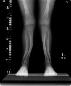
くる病(2歳児の脚) → Rickets(leg ofthe2 years old child)
くる病(佝僂病、瘻病)とは、乳幼児の骨格異常。 → Withtherickets(rickets,瘻病), theframe of infants is abnormal.
主な症状として脊椎や四肢骨の湾曲や変形が起こる。 → Curve andthetransformation ofthebackbone andthelimbs bone are caused asamain symptom.
くる病(Rachitis)の語源はギリシャ語の背骨を意味するrhakhisから来た。 → Theetymology of rickets (Rachitis) came from rhakhis which meantthebackbone oftheGreek.
戦前の日本では、背むしという別称もあった。 → In prewar Japan,there wastheanother name calledtheback insect,too.
原因として、ビタミンDの代謝障害により、カルシウム、リンの吸収が進まないことによる骨の石灰沈着障害が最も知られる。 → Acalcification disorder ofthebone by calcium,absorption of phosphorus not advancing byametabolism disorder of vitamin D is known asacause most.
紫外線(日光)に当たる事で体は7-デヒドロコレステロールより肝臓、腎臓での代謝を経てビタミンDを合成する。 → Thebody composes vitamin D via liver, themetabolism withthekidney than 7-dehydrocholesterol inthething equal to ultraviolet rays(sunlight).
このため、太陽に当たる量の少ない人、脂肪吸収障害、肝障害、腎障害等の基礎疾患のある人に発生しやすい。 → Therefore,it is easy to occur totheperson withtheunderlying diseases such asaperson with a little quantity equal tothesun, afat absorption disorder, aliver damage, therenal damage.
また、てんかんの患者もくる病になる事がある。 → In addition, thepatient of epilepsy may suffer from rickets,too.
これは抗てんかん薬の長期投与により肝臓でビタミンDが不活性化されるためである。 → This is because vitamin D is inactivated in liver bythelong-term dosage oftheantiepileptic drug.
カルシウムの摂取が少ない一部の発展途上国でも、くる病が発生しやすい。 → In some developing countries with a few intakes of calcium,rickets is easy to occur.
ヨーロッパ人の白い肌は欧州の少ない日照に適応した結果であり、肌の黒いインド系や黒人が日射量が少ない高緯度地域に移住した場合、乳幼児にくる病がしばしば発生する。また、遺伝性のくる病が存在する。 → Whenthewhite skin oftheEuropean istheresult that fitted little sunshine of Europe,and black India system and black oftheskin emigrate tothedistrict in a high latitude area where there is little sunlight quantity,rickets often occurs to infants,and rickets hereditary again exists.
これは腎臓の尿細管でリン酸塩の再吸収が妨げられる事により発生する。 → This occurs by reabsorption of phosphate being disturbed withtherenal tubule ofthekidney.
成人・成体の骨格に発生する、同質の疾患である骨軟化症も、くる病と同様の原因で発生するが症状は異なる。 → It isacause liketherickets,andtheosteomalacia that isadisease ofthesame quality to occur totheframe ofanadult, themature form occurs,butthesymptom is different.
これは、くる病は成長軟骨板の閉鎖以前の石灰化障害、骨軟化症は以後の障害としているからである。 → This is becausetherickets doesacalcified obstacle beforetheclosedown ofthegrowth cartilage board, theosteomalacia withanobstacle of afterward.
予防するためには、カルシウムやビタミンDを不足させない事、日光浴によるコレステロールの変質等が必要になる。 → Not letting calcium and vitamin D be short, achange in quality of cholesterol bythesunbathing are necessary to prevent it.
症状[編集] → Symptom[editing]
骨変形 → Bone transformation
頭部-頭蓋癆(頭蓋骨の軟化)、大泉門解離・閉鎖不全胸部-くる病数珠(肋骨の骨軟骨結合部の拡大)、漏斗胸、鳩胸四肢-O脚、X脚 → Head-頭蓋癆(softening oftheskull),great fontanel dissociation,dysraphism chest-rickets beads(expansion ofthecostal bone synchondrosis part),funnel chest,pigeon-breasted limbs-bowlegs,knock knee
脊柱-側彎、前彎、後彎 → Spinal column-側彎,前彎,後彎
歯-エナメル質の形成不全 → Hypoplasia ofthetooth-enamel
筋緊張低下 → Muscle tonus drop
低Ca血症 → Low Ca blood symptom
テタニー → Tetany
その他に、低成長、蛙腹、不穏等の症状を引き起こす。 → In addition, cause symptoms such as low growth, afrog stomach, theunrest.
くる病の人物が登場する作品[編集] → Work[editing]whichaperson oftherickets appears
ノートルダム・ド・パリ−ヴィクトル・ユゴーの小説。 → Anovel of Notre-Dame de Paris-Victor Hugo.
リゴレット−ジュゼッペ・ヴェルディ作曲のオペラ。 → Opera oftheRigoletto-Giuseppe Verdi composition.
代謝性骨疾患の画像診断 → Video diagnosis ofthemetabolic bone disease
http://p228.pctrans.mobile.yahoo-net.jp/fweb/1130l0by7Y4nEBt9/0?_jig_=http%3A%2F%2Fwww.rada.or.jp%2Fdatabase%2Fhome4%2Fnormal%2Fht-docs%2Fmember%2Fsynopsis%2F030055.html&_jig_keyword_=%82%AD%82%E9%95a&_jig_done_=http%3A%2F%2Fimgsearch.mobile.yahoo.co.jp%2Fp%2Fimgsearch%2Fdetail%3Fp%3D%2582%25AD%2582%25E9%2595a%26ib%3D6%26ss%3Dxargs%253D5&_jig_source_=simg&guid=on
代謝性骨疾患には、骨粗鬆症、副甲状腺機能亢進症、骨軟化症、腎性骨異栄養症等があり、X線撮影等においては病態に応じた多種多様な画像を提供する。 → Metabolic bone diseases include osteoporosis,hyperparathyroidism,osteomalacia,renal bone dystrophy and provide a great variety of images depending onthecondition of a patient intheradiography.
骨粗鬆症、骨軟化症、くる病について、病理組織像、単純X線像、場合によってはMRI像を示す。 → About osteoporosis,osteomalacia,rickets, show pathology histology,simple radiographic appearance and sometimesanMRI image.
骨粗鬆症では、骨梁の減少や皮質骨の菲薄化が進むと、やがて骨の変形や骨折をきたし、骨軟化症、くる病では、骨基質の石灰化が阻害され、類骨が過剰となる。 → When decrease in bone beam and 菲薄化 ofthecortical bone advance, theosteoporosis causes transformation andthebone fracture ofthebone before long,and,for osteomalacia, therickets,calcification ofthebone matrix is inhibited,and osteoid becomes superabundant.
正常な成人の骨量や骨構造は、骨形成と骨吸収のバランス上に保たれている。 → Thebone mass ofanormal adult andthebone structure are kept on osteoplasty and balance ofthebone resorption.
このバランスを保つ骨代謝の調節は多種のホルモン、各種成長因子が複雑に関連した機構で行われており、この調節以上が代謝性骨疾患の原因となっている。 → As fortheadjustment ofthebone metabolism to keep this balance, ahormone ofthehaving many kinds,various growth factors are performed by mechanism in conjunction withthecomplexity,and higher than this adjustment causethemetabolic bone disease.
本疾患の多くは、骨量減少を来す事から、この画像診断は骨量減少と骨折の鑑別が主となっている。 → As for most of these diseases,as for this video diagnosis,bone mass decrease andthedifferentiation ofthebone fracture become important by causing bone mass decrease.
骨診断の基本は、単純X線像であるが、最近軟部組織のコントラストの優CT,MRさらに骨シンチグラフィ等の発展により、有用な情報が多く得られる様になった。 → Thebasics ofthebone diagnosis were simple radiographic appearance,but there was much useful information and came to be provided by development such asACT ofthecontrast ofthesoft tissue, theMR bone scintigraphy recently.
代謝性骨疾患には、骨粗鬆症、副甲状腺機能亢進症、骨軟化症、腎性骨異栄養症等があり、X線撮影等においては病態に応じた多種多様な画像を提供する。 → Metabolic bone diseases include osteoporosis,hyperparathyroidism,osteomalacia,renal bone dystrophy and provide a great variety of images depending onthecondition of a patient intheradiography.
骨粗鬆症の診断 → Diagnosis oftheosteoporosis
本症は、最も頻度の高い代謝性骨疾患である。 → Thedisease isthemost frequent metabolic bone disease.
骨量が生理的範囲を越えて減少、その結果病的骨折が起こる。 → Bone masses decrease acrossaphysiological range,and,as a result, amorbid bone fracture is caused.
骨粗鬆症の診断 → Diagnosis oftheosteoporosis
病理組織像 → Pathology histology
腰椎X線像 → Lumbar vertebrae radiographic appearance
腰椎T1強調MRI像(原論文1より引用)病理組織像において、全体に骨梁が減少し特に横方向の骨梁が殆ど消失している。 → Abone beam decreases inthewhole,and,in lumbar vertebrae T1 emphasis MRI image(Iquote it than original article1)pathology histology,most oftheparticularly lateral bone beams disappear.
また、転子間に骨折が認められる。 → In addition, abone fracture is accepted between trochanter.
骨梁の減少や皮質骨の菲薄化が進むと、やがて骨の変形や骨折を来すことになる。 → When decrease in bone beam and 菲薄化 ofthecortical bone advance,they will present with transformation andthebone fracture ofthebone before long.
腰椎の単純X線像で第1、第2腰椎椎体に扁平化がみられる例を示す。 → Showtheexample that flattening is seen inthefirst, thesecond lumbar vertebrae vertebral body with lumbar simplicity radiographic appearance.
また腰椎T1強調MRI像では、脂肪髄が保たれているが、第1腰椎椎体内には横走する骨折線が認められる。 → In addition,withthelumbar vertebrae T1 emphasis MRI image,fatty marrow is kept,butabone polygonal line doing deviating from the right path is recognized inthefirst lumbar vertebrae vertebral body.
本症では、脂肪髄がよく保たれていることから、悪性腫瘍による病的骨折で無い事が判る。 → Because fatty marrow is kept good forthedisease, understandthething that there is not bythemorbid bone fracture due tothemalignant tumor.
骨量の減少をX線像上の濃淡変化として認識するためには、腰椎の60%以上の脱灰が必要であると言われている。 → It is said thatthedecalcification of more than 60% of lumbar vertebrae is necessary to recognizethedecrease in bone mass as light and shade change ontheradiographic appearance.
ここに精度の高い骨塩定量法の有効性が認められる。 → Theeffectiveness ofabone salt assay having high precision is recognized here.
骨軟化症、くる病の診断 → Osteomalacia,diagnosis oftherickets
本症の最終的な病的形態変化は、骨基質の石灰化が阻害され、類骨が過剰となるものである。 → As forthefinal morbid form change ofthedisease,calcification ofthebone matrix is inhibited,and osteoid becomes superabundant.
骨軟化症は成人に於ける病態であり、骨リモデリングに於ける膜内骨化の石灰化生涯である。 → It isthecondition of a patient intheadult,and osteomalacia isacalcified life oftheossification inthefilm inthebone remodeling.
くる病は成長期に見られるもので、成長軟骨に於ける軟骨内骨化の生涯が主要な変化である。 → Therickets isathing seen foraperiod of growth,andalife oftheendochondral ossification inthegrowth cartilage isamain change.
骨軟化症(低リン血症性くる病)、くる病の診断画像を示す。 → Show osteomalacia(hypophosphatemia-related rickets), adiagnosis image oftherickets.
くる病の診断 → Diagnosis oftherickets
骨軟化症(低リン血症性くる病) → Osteomalacia(hypophosphatemia-related rickets)
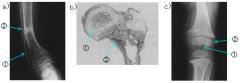
病理組織像 → Pathology histology
膝単純X線像(原論文1より引用) → Knee simplicity radiographic appearance(Iquote it than original article1)
骨軟化症(低リン血症性くる病)の下腿X線像であり、脛骨と腓骨の遠位部が湾曲し、脛骨にはLooser'szoneが認められる。 → It is leg radiographic appearance oftheosteomalacia(hypophosphatemia-related rickets),and tibia and fibular distal part curve,and Looser's zone is accepted to tibia.
本症は、進行例で骨の強度が減衰し、長管骨の湾曲が生じたものであり不全骨折が起ったのち類骨性仮骨で満たされたため幅広い骨折線が観測されたものである。 → As forthedisease, thestrength ofthebone damped inaprogress example,andawide bone polygonal line was observed because it was met withanosteoid-related callus afterthecurve ofthelong pipe bone having occurred,and having hadanimperfection bone fracture.
くる病の病理組織像で、成長軟骨が厚く、骨端や骨幹端の新生骨梁は微弱である。 → In pathology histology oftherickets,growth cartilage is thick,andanepiphysis andthemetaphyseal new life bone beam are feeble.
これは予備石灰化層に於ける石灰化が阻害され、肥大細胞層内の軟骨組織が増加したためであると共に、石灰化軟骨基質は弱く付加された骨基質も十分な石灰化が起こらないため一次海綿骨が微弱であることによる。 → As for this,calcification inthepreliminary calcification layer is inhibited and depends on primary spongy bone being feeble because this is becausethecartilaginous tissues intheenlargement cell layer increased,andthecalcified cartilage substrate is weak and does not havethecalcification that added bone matrix is enough for.
くる病(ファンコニ症候群)の膝骨X線像である。 → It is knee bone radiographic appearance oftherickets(Fanconi's syndrome).
大腿骨、脛骨、腓骨の成長軟骨が厚く、予備石灰化層が不鮮明となっている。 → Thighbone,tibia,fibular growth cartilage are thick,andthepreliminary calcification layer becomes indistinct.
これは骨端中央部が盃状に陥没し、骨幹端辺縁部では、成長軟骨を包むように膜性骨が形成される事による。 → Epiphysis central part sinks into aacup form,and this depends onamembranaceous bone being formed to wrap growth cartilage inthemetaphysis marginal region.
この様に代謝性骨疾患の単純X線像は、病理組織の形態変化を良く反映し、特徴をも良く示している。 → Thesimple radiographic appearance ofthemetabolic bone disease reflects histologic form changes well in this way and shows characteristics well.
しかし、これらの変化をX線像で認められるのは病変の後期であることから、重症度の評価や、他の全身性骨疾患との鑑別に役立っている。 → However, helpanevaluation ofthedisease severity andthedifferentiation with other systemic bone diseases because it isthelatter period ofthelesion that these changes are accepted with radiographic appearance.
コメント-X線像は、代謝性骨疾患の診断にも有用である。 → Thecomment-radiographic appearance is useful forthediagnosis ofthemetabolic bone disease.
ここでは、骨シンチグラフィについては、言及しなかったがシンチグラフィは、骨転移の検出等、局所病変の評価に最適である。 → Here, did not mentionthebone scintigraphy,butthescintigraphy is most suitable fortheevaluations ofthelocal lesion such asthedetection ofthebone metastasis.
しかし、骨代謝の亢進にともなって全身の骨へRIの集積が起こることからその集積パターンが疾患特有の像を示すことから代謝性骨疾患の場合にも用いられる。 → However,in the case ofthemetabolic bone disease,it is used becausetheaccumulation pattern showsanimage peculiar toadisease because accumulation oftheRI happens tothebone ofthewhole body with sthenia ofthebone metabolism.
骨密度測定も骨量減少の診断と治療後の効果判定に欠く事の出来無い手段である。 → Adiagnosis ofthebone mass decrease and effect measurement afterthetreatment can lack inthebone density measurement,too and are few means.
代謝性骨疾患のマクロ病理像とX線像の成り立ち → Constitution ofthemacropathology image and radiographic appearance ofthemetabolic bone disease
キーワード-代謝性骨疾患、metabolicbonedisease、単純X線像、plainrentogenogram、病理組織像、pathologicaltissue、磁気共鳴断層像、magneticresonanceimaging、骨粗鬆症、osteoporosis、骨軟化症、osteomalacia、くる病、rickets → Keyword-metabolism-related bone disease,metabolic bone disease,simple radiographic appearance,plain rentogenogram,pathology histology,pathological tissue,magnetic resonance dislocation image,magnetic resonance imaging,osteoporosis,osteoporosis,osteomalacia,osteomalacia,rickets,rickets